


When discussing clinical equipment and personal protective equipment, it is vital to appreciate the difference between a ventilator and a respirator. Although these words are sometimes considered synonyms, they denote different devices tailored for particular uses within the contexts of healthcare and safety. This article seeks to explain such differences by discussing functions, designs, uses, and more. From medical experts and safety engineers to laypersons seeking to gain more knowledge about these essential instruments, this guide is meant to provide everyone with an easy yet informative understanding of these tools, their functions, and importance.
What is a ventilator, and how does it work?
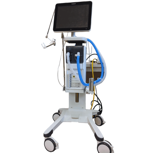
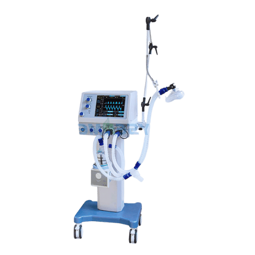
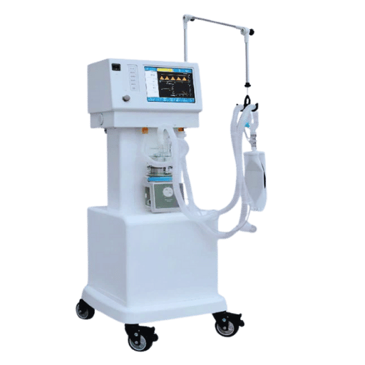
The medical apparatus that aids or replaces the breathing process for individuals unable to breathe without assistance is called a ventilator. It works by providing oxygen as well as removing carbon dioxide to the lungs, thereby ensuring effective gas exchange. A ventilator can be set to fully control breathing or to support a patient’s attempts at breathing. These devices are used in emergency hospital units or intensive care units, during surgical procedures, or in patients with respiratory diseases resulting in reduced lung function. The primary goal of a ventilator is to retain an acceptable concentration of oxygen and avert hypoxic respiratory failure.
How does a ventilator help with breathing?
A ventilator helps in breathing by providing a controlled flow of oxygen-rich air to the lungs, using either invasive methods like endotracheal intubation or non-invasive means like a mask. Modern ventilators are intricately sophisticated, offering precise control over air pressure, volume, and flow rate for tailored ventilatory support. They can also adjust these values in real-time while taking into account functions like oxygen saturation, respiratory rate, and tidal volume, anticipating optimal lung function.
Evidence suggests that ventilators are particularly useful in mitigating the impacts of ARDS, a severe ailment that may accompany other pneumonia or COVID-19-related critical illnesses. Research indicates that certain ventilation methods, which use lower tidal volumes with pressure limits, have better survival rates and less lung damage. Moreover, ventilators are fundamental to offloading the respiratory muscle workload for patients in critical care. These units, along with many others, allow the body to recuperate by optimizing healing processes, proving crucial in fighting respiratory failure. They rectify relevant oxygenation and carbon dioxide clearance in life-threatening medical situations.
What are the components of a mechanical ventilator?
A mechanical ventilator, or just ventilator as it is often referred to in hospital settings, is a sophisticated piece of equipment with multiple functions crucial to helping a patient’s breathing processes. The key functions of a ventilator include:
Gas Supply System
The ventilators main functions is agility in pumping measured and medically mixed oxygen, oxygen and ehriled air along with other components needed to make a patient’s breath clean, compounds coming out are sucked out via flow and pressure sensors, giving exremely accurate measures alongside gasses.
User Interface
The display has multi PLCs which let the hypermodernized machines set ergometers PcontP, P pressure editorial ventilation – PL tidal volume, RR, I time to E, PEEP. The newest models have a interfaces that moves with users finger and dictate commands to computer help programs.
Breathing Circuit
The hybrid ventilator’s main frame connects to the rest of the machine with tubes tied to parts of the body using an endotracheal or tracheostomy tube. The sucg system makes it possible for an admixture of gases to get into the lungs and allow the removal of used eluanted gases. It includes inspiratory parts and expiratory limbs, one of the newly added legge to other body parts, Hoch-dryer, and new filters and pressure-regulated valves.
Ventilator Modes and Software
Among the different modes of operation in ventilators, volume-controlled, pressure-controlled, and spontaneous modes are available in varying degrees to suit the requirements of the patient. Breathing software has been advanced to improve the efficiency of the ventilation process, monitor respiratory mechanics, and visualize data and metrics in real-time.
Pressure and Flow Monitoring Systems
Monitoring Airway pressure, tidal volumes, and flow rate is continuously measured through the ventilator’s sensors. These measurements are useful in the adjustment of parameters as well as patient safety by mitigating risks like barotrauma and autotrapping which occurs when ventilation parameters improperly set.
Exhalation Valve
The exhalation valve permits the free passage of exhaled air while setting a limit to the ingress of air during the breath-holding periods at pre-specified PEEP levels. It ensures the setting pressures prevent alveolar collapse.
Alarm System
Safety alarm systems monitor clinical parameters such as airway pressure, tidal volume, and apnea as set parameters within set ranges. Alarms are systemized to enhance the safety levels by reducing the risk of dangerous situations by enabling immediate response.
Humidification System
Ventilators deliver gas in dry form, which makes the inclusion of a humidifier crucial. Newer devices incorporate other components such as weight, height, and moisture exchangers to improve the quantity and quality of ventilatory gas.
Mechanical ventilators enhance patient outcomes with sophisticated hardware alongside advanced algorithms, improving respiratory support in critical scenarios with precision and control.
When might someone need a ventilator?
A patient may require a ventilator when breathing is inadequate due to either acute or chronic conditions. For instance, acute respiratory distress syndrome (ARDS) is one of the most prevalent causes of ventilator dependency, impacting around 10% of ICU patients worldwide. Other medical emergencies, such as pneumonia, severe asthma, or chronic obstructive pulmonary disease (COPD), also compromise respiratory function and require mechanical ventilation.
In surgery, ventilators are critical during periods of general anesthesia where breathing is inhibited, allowing patients to receive adequate oxygen during the entire procedure. Moreover, people with neuromuscular diseases such as amyotrophic lateral sclerosis (ALS) or spinal cord injuries may need ventilation aid because their muscle strength fails them. Emerging evidence underlines the need for ventilators during pandemics. Acute Respiratory Distress Syndrome has been coupled with the COVID-19 pandemic, severely impacting cases with respiratory distress. Studies reported that 2.3% of hospitalized COVID-19 patients required mechanical assistance to breathe, illustrating the surge in ventilator dependency.
In summary, the use of ventilators is fundamental when dealing with critical matters such as ensuring proper oxygen supply and required medical attention when respiratory functions are weakened in patients.
How does a respirator differ from a ventilator?
What is the primary function of a respirator?
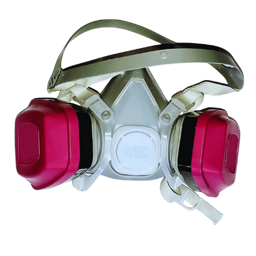
The principal purpose of a respirator is to prevent a person from breathing in harmful, airborne contaminants such as particles, gases, or vapors. As NIOSH outlines, PPE includes disinfecting equipment (like respirators) that filter inhalable contaminants away from the air that the user breathes. For instance, N95 respirators, which are widely utilized in industrial and healthcare settings, are designed to block no less than 95% of very tiny (0.3 microns) particulate matter. These devices are essential in settings such as construction sites, laboratories, or during outbreaks of infectious diseases, where there is a high risk of exposure to hazardous substances. Research shows voluntary use of certified respirators greatly decreases the chances of contracting airborne pathogenic diseases and enhances safety in both work and personal environments.
How do respirators protect the wearer?
Respirators safeguard the user by forming a sealed barrier between the respiratory system and dangerous airborne contaminants. This protection is achieved through a combination of filtration and proper fit. Particulate filters in respirators, such as HEPA filters, can capture particulates as small as 0.3 microns with no less than 99.97% efficiency. Another example is N95 respirators, which are designed to filter out 95 percent of particulates, including dust, smoke, and allergens, as well as bacteria and viruses.
To achieve maximum protection, respirators rely on the material and proper fit. Air filtration ensures that hazardous air particles are removed before inhalation, while the respirator’s design ensures unfiltered air cannot circumvent the mask. Research furthermore proves that properly fitted respirators afford substantial protection against airborne contaminants, including droplets with infective agents. One example is the use of N95 respirators by healthcare workers, which greatly reduced infection risk, a critical protective measure in high-risk environments.
Moreover, respirators undergo testing, wear, or are evaluated to ensure their proper functionality under harsh conditions. One of the most rigorous tests includes the National Institute for Occupational Safety and Health (NIOSH) certification for the United States. Rest assured, these devices will function properly in industrial, medical, and even emergency. Proper training on putting on a respirator and conducting fit-test verification increases the effectiveness of the protection provided by the device.
In what situations are respirators typically used?
Respirators are most often deployed in environments with hazardous airborne contaminants or where oxygen is scarce. These environments encompass industrial work with dangerous dust, fumes, or chemicals, like construction, mining, and manufacturing. They are also used in healthcare settings for protection against infectious biological agents during outbreaks of airborne diseases. Furthermore, emergency responders use respirators in situations involving hazardous material spills, fire, or rescue operations in oxygen-deficient atmospheres.
What are the different types of ventilation?
How does non-invasive ventilation differ from invasive ventilation?
The main distinction between non-invasive ventilation (NIV) and invasive ventilation lies in the method of support delivery. NIV maintains respiratory support externally using masks or nasal prongs and does not require any type of surgical intervention. Invasive ventilation, however, requires direct access to the airway via a tube inserted through the mouth (intubation) or surgically placed in the neck (tracheostomy) for direct lung access. While invasive ventilation is generally utilized for critical and prolonged life support, NIV is more commonly used in diagnostically milder cases or as a first step to preempt the need for intubation.
What is the role of CPAP and BiPAP in respiratory care?
Both CPAP (Continuous Positive Airway Pressure) and BiPAP (Bilevel Positive Airway Pressure) are examples of non-invasive ventilation used in the clinical setting. This technique is characterized by offering a seamless, continual stream of air pressure that prevents the airway from collapsing. This makes CPAP effective in the treatment of obstructive sleep apnea diseases as well as helping to diminish the energy expended on breathing. BiPAP, on the other hand, provides two different levels of pressure: a higher pressure for inhalation and a lower pressure for exhalation. This is useful for people with chronic obstructive pulmonary disease (COPD) or respiratory failure. All these procedures help in the improvement of oxygen levels, aid in ventilatory effort, and decrease the need for more invasive procedures.
When is a breathing tube necessary?
An endotracheal or breathing tube is needed in medical situations where a patient is unable to independently maintain an adequate airway or proper oxygenation on their own. This life-sustaining intervention is often needed during medical crises, surgical operations under general anesthesia, or acute respiratory distress. Indications of this intervention involve acute respiratory failure, trauma to the airway, severe allergic response with airway obstruction, and status asthmaticus refractory to other therapeutic interventions.
Data suggest that intubation is a common practice performed on patients admitted to intensive care units. Around 30–40% of critically ill patients require mechanical ventilation. Action and prompt decision-making are essential; in fact, studies suggest that delaying intubation in the setting of worsening respiratory function greatly increases mortality. For example, clinical guidelines suggest di-intubation when oxygen saturation is less than 90% on two liters of oxygen or hypercapnia (high carbon dioxide levels) on blood gas tests hypercapnia confirming the need for intervention. Additionally, intubation is vital to protect the airway during effective ventilation and evacuation of air for non-breathing patients in cardiac or respiratory arrest.
As this procedure is very technical, it must only be undertaken by a qualified medical professional to reduce the risk of complications like inadvertent esophageal intubation, airway damage, or infections. Evaluation and close observation are critical to assess the level of intubation as well as the timing for extubation or prolonged mechanical ventilation, dependent on the patient’s recuperation and health status.
How does a breathing tube assist in ventilation?

What is the purpose of an endotracheal tube?
An endotracheal tube (ETT) is a medical device which ensures that an airway is unobstructed for the inflow oxygen and anesthetic gas to the lungs while serving as a facilitator in the efflux of carbon (IV) oxide gas. The aim of endotracheal tube is to assist mechanical ventilation on patients who are insufficiently breathing because of a medical condition, injury, or during surgical operations requiring general anesthesia.
The tube is made of pliable, biocompatible polyvinyl chloride or silicone and is inserted through the mouth or nose into the trachea. Once positioned correctly, a cuff inflated at the distal end of the tube creates a seal that prevents air leakage and the aspiration of contoured gas mixed with gastric content into the lungs. An ETT, when properly managed, has been shown to enhance oxygenation and ventilation during critical care, especially with ARDS, improving survival rates when intubation is done timely and is managed well.
Moreover, the tube comes fitted with a connector at the proximal end for integration with ventilators, facilitating control of tidal volumes, respiratory rate, and oxygen concentration for each patient. Modern ETTs often include such features as radiopaque markers to facilitate imaging, and subglottic suction to reduce the risk of VAP (ventilator-associated pneumonia), a complication that affects as many as 20% of patients undergoing mechanical ventilation.
Endotracheal tubes remain one of the most important components of advanced airway management, and they provide crucial assistance in emergencies, during surgical procedures, and in the intensive care unit (ICU), often under intricate and life-threatening situations a person may encounter.
How is a tracheostomy performed?
A tracheostomy is a surgical procedure that creates an incision in the neck to directly access the trachea (windpipe). The surgical guide includes the steps below:
- Preparation: Identify the patient’s position with an extended neck and apply antiseptic preparation to the neck. Follow with a general or local anesthetic to eliminate any discomfort raised by the procedure.
- Incision: Execute a small horizontal or vertical cut on the inferior neck above the sternum.
- Accessing the Trachea: After all tissues are separated, identify the trachea and perform a cricothyrotomy by taking excision of tracheal wall so as to create external airway (stoma).
- Tube Placement: Fix a tracheostomy tube in the aperture to provide direct access for respiration.
- Securing the Tube: Tie the suture of the tube to the neck by means of other loose straps to prevent anchoring dislocation.
The last part of the surgical work includes ensuring the tube checked to works properly, while also observing the patient on a <specific buzzword for monitor> for irremediable trends for everything. The best resolution for breathing problems is the cut done organically for patients suffering from asthma attacks.
What are the risks of using a breathing tube?
While using a breathing tube may be somewhat of a necessity, it comes with considerations as follows:
- Infections: If a breathing tube is used for a longer time, then patients become more susceptible to infections like pneumonia.
- Airway Damage: The tube may also lead to some form of irritation, injury, or scarring in the throat or wind pipe.
- Blockages: There may be blockages due to mucus or other materials, which may limit airflow.
- Difficulty Weaning: After prolonged use, patients struggle with trying to adapt to life without the tube.
- Discomfort: Breathing tubes may lead to discomfort such as coughing, gagging, or pain in the throat.
Appropriate observation is very important in order to reduce risks along with appropriate care so that patient safety is maintained.
What are the challenges of using a face mask for ventilation?

How do face masks deliver positive airway pressure?
Face masks ensure that pressurized air enters a patient’s airway by delivering positive airway pressure through a sealed interface over the nose and/or mouth. This occurs with CPAP and BiPAP machines, which function as non-invasive ventilation (NIV) devices. CPAP systems are designed to constantly and steadily deliver air and are commonly used in the treatment of obstructive sleep apnea. BiPAP systems offer tailored support by alternating between higher and lower pressures for patients with more complex breathing disturbances.
As with all medical devices, the system’s effectiveness relies on a few critical aspects: proper fitting of the face mask, suitable interface pressure, and the patient’s respiratory patterns. Studies indicate that moderate to severe sleep apnea patients can achieve a 70% reduction in apnea-hypopnea index (AHI) levels with CPAP therapy, greatly enhancing oxygen saturation while decreasing cardiovascular strain. Modern face masks also minimize air leakage thanks to advanced materials combined with ergonomically designed features, which provide patient comfort while combating skin irritation and pressure sores.
Positive pressure mask ventilation is an integral aspect of respiratory therapy in intensive care or anesthetic settings where oxygenation and ventilation must be meticulously controlled. Technology such as adaptive pressure systems and real-time monitoring is advancing the accuracy and effectiveness of positive airway pressure delivery through face masks.
What are the common issues faced with face masks?
Though face masks have an important function in respiratory therapy, their application is often linked to numerous problems. One major concern is air leakage, which can impair ventilation effectiveness and worsen the patient’s overall condition. Some research indicates leakage rates of up to 40% in certain situations, particularly with inadequate mask fit or problematic facial structure.
Skin problems are also common. These include pressure ulcers, skin redness, or skin irritation from prolonged mask use. Research estimates that these problems occur in more than 30% of the patients who are on long-term masking therapy. The problems can strain patients and in extreme instances, lead to noncompliance with treatment.
Inadequate humidification of air that is channeled through the mask also leads to airway dryness and nasal congestion, which are common complaints. This tends to worsen a person’s already delicate respiratory condition.
Furthermore, health compliance is something to pay attention to. Reduced adherence due to discomfort from poorly fitting masks, ventilator noise, or anxiety about wearing a mask can be detrimental to therapeutic results. For instance, research indicates that adherence rates for patients undergoing Continuous Positive Airway Pressure (CPAP) therapy are estimated to reach a mere 50% because of these factors.
Last but not least, some of the challenges above are being aided by new mask designs with advanced cushioning, adaptable fittings, and other features. Regardless, these persistent problems still require ongoing refinement through Active engagement, education, and tailored interventions with the patient.
How do healthcare professionals manage ventilator support?

What role do respiratory therapists play?
Respiratory Therapists (RTs) specialize in ensuring proper respiratory management of patients on mechanical ventilation, overseeing active ventilator support, and coordinating multidisciplinary care about the patient’s ventilation needs. They assess the patient’s respiratory condition, set the ventilator parameters, and supervise complications such as ventilator-induced lung injury (VILI) and oxygen toxicity.
Among these duties, RTs need to perform routine assessments of the ventilator-measured parameters such as tidal volume, breaths per minute, and PEEP, ensuring optimal lung mechanics and oxygen delivery calculations. Studies show RT-led initiatives substantially improve complication rates and overall ventilation outcomes alongside patient comfort. For instance, more protective ventilatory strategies such as low tidal volume ventilation have been demonstrated to reduce mortality in ARDS patients. Moreover, RTs train and educate caregivers and patients regarding the use of home ventilation systems, ensuring ongoing care after active medical supervision ends.
RTs’ technical knowledge with a patient-first care approach enables them to directly address issues within patient specific healthcare needs, providing tailored solutions, thus greatly improving the quality of care in critical conditions.
How is the respiratory system monitored during ventilation?
Monitoring the effectiveness of the respiratory system during the process of mechanical ventilation requires continuous assessment in order to ensure optimal safety, support, and complication management. Ensuring each set parameter is continuously optimized requires the integration of multiple modern techniques and technologies by the clinician.
- Airway Pressure Monitoring: Monitoring Airway Pressure requires measuring the resistance in the patient’s airway, as well as, measuring the compliance of the lungs during the process of mechanical ventilation. Errors in range displayed by airway pressure monitoring devices suggest poor settings of the airflow machines, blockages in the airways, or loss of lung function.
- Tidal Volume and Minute Ventilation: In addition to tracking time per breath, tidal volume refers to the volume of ventilated gas in the lungs per time interval and, as such, assists in determining ventilatory support. As such, bolus over-distension of the lungs as well as over-saturation may occur.
- End-Tidal CO2 (ETCO2) Monitoring: This parameter, being the amount of carbon dioxide released when the patients cease to breath, is essential in evaluating the ventilation schedule. Conforming values exist but do not exceed 35-45 mmHg value and changes are associated with hypoventilation, hyperventilation, or other physiological changes.
Oxygenation Measurements:
- While oxygenation measures the infused oxygen within the body, this one works without touching the person. Detecting SpO2 also makes it easier to gauge operational performance and even detect hypoxemia.
- Arterial Blood Gas (ABG) Analysis: Measured metrics from ABG tests, including the partial pressures of oxygen and carbon dioxide (PaO₂ and PaCO₂, respectively) and pH, provide insights into a patient’s oxygenation, ventilation, and acid-base balance.
Lung Mechanics and Compliance:
Dynamic Compliance (Cdyn) and Static Compliance (Cstat) are two metrics acceded to measure lung resistance and elasticity. Reduced compliance might be indicative of acute respiratory distress syndrome (ARDS) or pulmonary fibrosis.
Monitoring of Peak Inspiratory Pressure (PIP) and Plateau Pressure (PPLAT) is important to prevent hyperbaric barotrauma injury from excessive ventilation pressure.
The patient’s respiratory rate needs to be checked concerning their specific physiological range, so synchrony within the ventilator-patient interaction must be gauged to avoid significant gas exchange inefficiencies.
Advanced techniques like EIT offer real-time, non-invasive imagery of the lung ventilation region, enabling the clinician to adjust settings and avert complications like VILI.
Through applying these techniques, the patient, during mechanical ventilation, has better outcomes alongside enhanced clinical decision-making deriving from more tailored interventions.
What are the signs that a patient can be removed from ventilator support?
Weaning, the process of assessing if a patient can safely be removed from mechanical ventilator support, involves careful consideration of the patient’s clinical and physiological assessments as well as the psychological readiness caps. Predominantly, the following indicators signal readiness for extubation:
1. Respiratory Status Externally Observable:
The stable respiratory status of the patient should be characterized by even chest rise and fall, respiratory movement shallows below 35 and tidal volume is above 5 shared per kg of the anticipated body weight.
2. Oxygenation Advanced Provision Criteria:
An ideal partial pressure of oxygen (PaO2) suggests adequate oxygenation when its value is above 60 mm Hg with a fraction of inspired oxygen (FiO2) not exceeding 0.4 – 0.5, in addition to a positive end-expiratory pressure (PEEP) that is below 5 – 8 cm H20. Moreover, the P/F ratio (PaO2/FiO2) should optimally be greater than 150-200.
3. Acid–base balance should be within the required bounds.
Failure or success of a patient to explicitly maintain adequate gas exchange by above venting or below venting strongly anchors a patient in the range of 7.35-7.45 without significant hyper/hypo-ventilation respectively. To expect gentle pH change, one has to look at arterial blood gas (ABG) analysis and observe breathing switch up or down .
4. Stabilization of hemodynamics:
Cardiovascular features shall not undergo major changes, pointing to vasopressor dependency as well as having no arrhythmias and hypotension, or any of the hemodynamic instability matters.
5. Adequate Neurological Status
Damage to the airway protective mechanisms or the failure to provide appropriate levels of cognition and consciousness protective mechanisms requires a GCS 8 or above or command compliance in following tasks.
6. Successful Spontaneous Breathing Trial (SBT)
Assessment of ventilator independence is best achieved with SBT. In an SBT, the patient breathes without assistance for 30-120 minutes, usually through a T-piece, and the clinicians look for signs of fatigue and increased work of breathing. During this time, the patients are monitored closely for signs of exhaustion, increased breathing rate (tachypnea), or decreased oxygen levels (hypoxemia).
Research Data on Extubation Success Rates
Recent clinical studies reveal that adherence to these parameters significantly improves extubation success rates. Planned extubation success rates are between 70 and 80 percent if the criteria for readiness are followed. Attempts to prematurely extubation increase the likelihood of needing to reinsert the tube, which has a high correlation to morbidity and mortality and prolonged stays in the hospital. Multidisciplinary evaluations for the most uncomplicated delivering patient care shift, while the patient shifts from relying on a ventilator to manual ventilation for optimal patient outcomes, are essential.
Frequently Asked Questions (FAQs)
Q: What is a ventilator?
A: A ventilator is a machine that directly provides oxygen to the lungs, which actively assists the user in altering the oxygen and carbon dioxide gas levels in the body. The use of this equipment is common in patients with advanced stages of COVID-19 or other transitional breathing issues.
Q: How does a respirator differ from a ventilator?
A: A respirator is a mask worn on the face that protects the nose, mouth, and lungs from inhalation of poisonous substances. Unlike spectrometry, a ventilator actively aids, and in some cases, completely takes over the breathing process of a patient.
Q: When is a patient typically connected to a ventilator?
A: Patients are connected to a ventilator when they are struggling to put enough oxygen into the bloodstream. This is common with acute respiratory diseases, during surgery with general anesthesia, or when a COVID-19 illness shatters lung capabilities.
Q: Can a ventilator be used noninvasively?
A: Yes, through the application of a mask or similar devices that encompass the nose and mouth, noninvasive ventilation can be administered. Such devices are capable of providing additional oxygen and maintaining the patency of airways without the need for intubation.
Q: What is intubation, and why is it necessary?
A: Intubation is the process of passing a tube through the nose or mouth into the trachea to secure an open airway in conjunction with mechanical ventilation. Intubation is necessary when the patient does not possess adequate respiratory effort and requires mechanical assistance to increase the oxygen levels in the lungs.
Q: How does a ventilator help in cases of COVID-19?
A: A ventilator aids in the sustained delivery of oxygen to the bloodstream. COVID-19 can severely damage lung function in addition to the normal oxygen and carbon dioxide exchange.
Q: How important are blood tests during the oxygenation of a patient with a ventilator?
A: Blood work assesses the oxygen and carbon dioxide content of the blood, so modified ventilator settings are tailored to the patient’s needs while confirming that the lung function, alongside the patient’s needs, is optimal.
Q: Is it possible for a ventilator to have consequences on the vocal cords?
A: Prolonged intubation is likely to impact the vocal cords as the tube moves through the laryngeal area. This region is at risk, so the non-invasive IPPV is preferred to reduce risk and needs stringent surveillance.
Q: What is the process of using a ventilator to remove carbon dioxide from the lungs?
A: During mechanical breathing either fully or partly supported by the ventilator, CO2 is removed alongside lung inflation and its sufficient elimation while oxygen delivery ensures continual adequate blood organ perfusion.
Q: Why are the airways maintained open in the case of a patient on ventilator support?
A: It is critical for ensuring that air can freely flow in and out for the lungs to facilitate the oxygen and carbon dioxide exchange, requirements which serves the optimized functioning of vital organs as well as overall well-being.
Reference Sources
1. Learning Effects of Incorporating XR Simulators for Mechanical Ventilation and Tracheal Suctioning Into a Novel Simulator (2024) (“Learning Effects of Incorporating XR Simulators for Mechanical Ventilation and Tracheal Suctioning Into a Novel Simulator,” 2024)
Key findings:
- The training task ‘endotracheal suctioning for a patient on a mechanical ventilator’ indicated a lower threshold of difficulty that was mastered during the 3rd grade and optimal for the 4th grade.
- Principal component analysis reveals two principal components: “Balance between learning content difficulty and motivation (Achievability)” as well as “Balance between the learning time/cost and the learning effect (Feasibility)”.
Methodology:
- The questionnaire was conducted post-lesson using Simmar+ESTE-SIM XR with fourth-year university students and faculty.
- Judged the learning effects of the simulator, as well as the criteria for judgment indicators when introducing the new simulator.
2. Development of Personal Protective Respirator in Healthcare: An Emergency Perspective (2023) (Selvakarthi et al., 2023, pp. 686-690)
Insights of the Work:
- For the purpose of coming up with a novel approach to the flexible and functional ventilator, Arduino was employed and the model comprises a silicon respiratory pack, a servo motor, and a lateral push actuator.
Technique of Work:
- A dependable yet inexpensive ventilator aid during pandemic scenarios was also built using Arduino; thus, the ventilator met the requirements.
3. EVALUATING A PATIENT’S READINESS TO BE WEANED OFF A RESPIRATOR FOLLOWING MECHANICAL LUNG VENTILATION(2023) Cherniaiev & Dubrov
Key Details:
- Predictive assessment of ventilator dependency in elderly dementia patients poses unique challenges due to the complex mental status evaluations, and the use of objective measures aids in the decision processes concerning the use of ventilators.
Methodology:
- Conducted SBT with inspiratory pressure support and ventilator dependency predictors, including Motor activity as rapid shallow breathing index (RSBI), diaphragm thickening fraction (DTf), and diaphragmatic excursion (DE) ratio were calculated.
4. Ventilator
5. Respirator
6. Breathing